If you’re dealing with frequent back or neck pain, your posture might be playing a bigger role than you think. Whether you're sitting at a desk all day or scrolling on your phone for hours,…
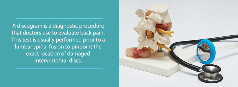
A discogram is a diagnostic imaging procedure that doctors use to evaluate back pain. Sometimes called discography, this method uses X-ray imaging to reveal abnormalities in the intervertebral discs of the spine. With the aid of a special dye and a fluoroscope, discograms are able to locate the cause of the patient’s back pain. The procedure may vary slightly between discographers, but the general purpose is always the same. It’s a diagnostic tool that doctors use to identify probable causes for a patient’s symptoms.
Do I Need a Discogram?
Medical experts consider discographies as an invasive test. Naturally, this means that it is not usually the first treatment that doctors use for evaluating the patient’s condition. In most cases, your doctor will try to treat your back pain using more conservative methods. This may include treatments such as physical therapy or medication. If these non-surgical techniques do not prevail, your doctor will then consider more drastic solutions.
Sometimes, when a patient needs lumbar spinal fusion surgery, the doctor on the case will perform a discogram before the procedure. The diagnostic test is useful here, as it reveals the affected discs in the spine beforehand. While this may be so, it is important to illustrate that discograms do not always provide accurate results. Some doctors instead prefer using other techniques, such as MRI and CT scans. The method that will be used for your specific case will depend on a variety of factors. It will depend on your doctor, your condition, your medical history, and you. Make sure that you communicate with your doctor if you have any preferences for any procedure.
A doctor may consider using a discogram if the patient has the following conditions:
- Generalized disc pain
- Degenerative disc disease
- Herniated disc
- Bulging disc
These are just a few general examples. Because discs act as the shock absorber between spinal bones, there are a lot of potential problems associated with them. Constant pressure and impact on these structures may lead to problems like the ones listed above.
Discogram Associated Risks
Discograms are quite safe, with complications happening only rarely. These risks are often minimized when the procedure is performed by a skilled specialist using modern discography methods. Although they are rare, the following complications may occur during this test:
- Allergic Reaction: Some patients are sensitive to the contrast dye used in the test, which results in an allergic reaction. Usually, this risk is averted by providing your doctor with an extensive medical history beforehand. Aside from reactions to the dye, there is also a chance for the patient to be allergic to anesthesia. You should communicate all medication allergies to your doctor before getting a discogram.
- Disc Space Infection: This is the most serious complication associated with discograms. Disc space infections are very rare but when they occur they are very hard to treat. Luckily, advances in sterilization have made this complication virtually nonexistent.
- Nerve Root Injury: Discograms inject the contrast dye into the affected area using a needle that passes quite close to nerve roots. Because the needle travels so close to these structures, there is a very small chance of nerve injury. This complication leads to additional pain that the patient feels after the procedure.
- Excessive Bleeding: Make sure to tell your doctor if you have any bleeding disorders before receiving any invasive treatment. Additionally, you should notify your doctor if you take any blood-thinning medications, such as aspirin. If you communicate with your doctor, the chances of this complication are very rare.
How is a Discogram Performed?
Firstly, the doctor must prepare the patient by using local anesthesia and a sedative. This will relax the patient and reduce uncomfortable sensations during the test. The doctor will then either lie the patient face down or on the side for access to the back. After the doctor sterilizes the injection site, the hollow needle is inserted where it is needed.
To make the injection go as smoothly as possible, a radiologist will often aid navigation using fluoroscopic guidance. Once the doctor determines that the needle is at the affected site, the contrast dye is injected. This may intensify uncomfortable symptoms, causing the patient some level of discomfort. Though this may be the case, it is important that the patient stays still for the procedure to be a success.
Finally, the doctor removes the needle and takes X-ray images of the affected site. Again, in order to obtain accurate images, it is vital that the patient stays still. In some cases, the doctor may perform a subsequent CT scan after the procedure.
Although the procedure is more invasive than conservative methods, it is still a minimally invasive technique. Additionally, discograms are also outpatient procedures, as the patient is able to go home the same day. Typically, the medical staff will observe the patient for 30 minutes directly after the test. Once the patient is home, medical experts recommend that he or she rest for the next 24 hours. Some patients may experience headaches after having a discogram. In these cases, the patient should take ibuprofen or Tylenol, but never aspirin.
Contact Us
If you have back pain that does not resolve on its own, please contact us at (855) 853-6542. At OLSS, you will find an efficient facility filled with caring spine doctors whose goal is to treat your ailments. Our doctors will take the time to understand the specifics of your case and they will come up with a treatment plan that best serves your needs.


