Back pain is incredibly common, and for many people it starts gradually. A sore lower back after a long day, stiffness in the morning, or discomfort after lifting something heavy can feel routine. But when…
The difference between x-rays, MRIs, and CT scans really boil down to what needs to be seen or what tissue is suspected to be causing a problem.
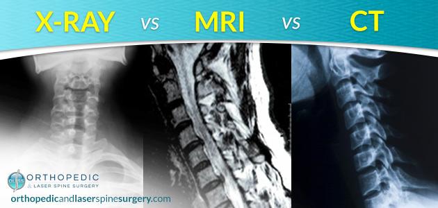
What Is an X-Ray?
The quickest radiological exam of the three is an X-ray. Most of us have had an x-ray whether it was at our dentist or when we fell off the jungle gym in elementary school. X-rays illuminate bone with one single “snapshot” image at a time. X-rays use ionizing radiation to create images of bones. Under x-ray, a bone glows white, and any break in that surface will be noticeable with a darkened area where there is a crack, break, or imperfection on the bone’s surface.
Therefore, X-rays are used frequently for traumatic bone injuries or to examine a bone deficit that would be easily detected under X-ray. An X-ray is generally not a sufficient radiological exam for tissue other than bone.
What Is an MRI?
MRI stands for Magnetic Resonance Imaging. And it is just what it implies. It uses a magnetic field to produce images instead of using radiation. Similar to the CT, during an MRI, the patient lays on a flat surface while the magnetic field generates pulses of images through the area being examined, creating an orderly series for the doctor to view.
MRI images will show soft tissue such as organs, tendons, and ligaments, and it offers the best view of the spinal cord. MRI machines use radio waves and magnetic fields to create detailed images of soft tissues. MRI scans are particularly useful for imaging soft tissues such as muscles, tendons, and ligaments. Bone will not illuminate brightly with an MRI, and this allows for better dense imaging of organs such as the brain, which can see past the thick encapsulating skull bones that protect it.
An MRI can be used to diagnose many things, but it can be a neurologist or neurosurgeon’s best tool when it comes to looking into spinal cord injuries, infections or bleeding in the brain, and other central nervous system disorders.
What Is a CT Scan?
A CT scan, or “CAT scan,” is the big brother of the simple X-ray. A CT uses the same type of radiation as an x-ray but in a much more detailed way. While the patient lies on a flat surface, a CT scan will rotate independently around the patient and take multiple cross-sectional images. It is kind of like having a bird’s eye view of your body that slowly moves from the top of the tissue being viewed and through to the bottom via a series of images. These series of images are placed on the CT’s integrated computer, and the doctor is able to view each cross-sectional image in the order in which it was taken and look for imperfections as the series progresses.
CT scans use computed tomography to create cross-sectional images of the body. CT scans are a type of computed tomography scan. CT scans can create detailed images of bone structures and are useful in emergency situations due to their speed. CT scans use ionizing radiation, which can pose risks with repeated exposure. CT scans are a valuable imaging test for diagnosing acute conditions such as brain hemorrhages and fractures.
CT scans are one of the many imaging tests used in diagnostic imaging techniques. CT scans create cross-sectional images that are crucial for accurate diagnosis and treatment planning. CT scans can create detailed images of internal organs and structures.
Of these three radiology options, spine specialists like those at Orthopedic and Laser Spine Surgery, feel CT Scans are the most beneficial for their patients with spine concerns. A CT illuminates the vertebral bones brightly, gives clear views of the discs and other supportive structures, and offers important details with the cross section viewing.
Contact Us Today!